Phospho Explorer Antibody Array
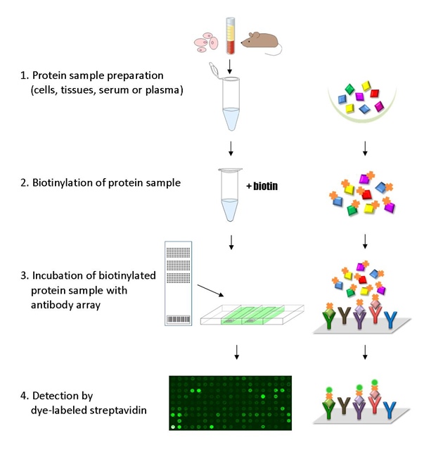
Protein phosphorylation plays an important role in cell signaling and metabolism. Phospho Explorer Antibody Array serves as a high-throughput, ELISA based, phosphorylation assay for qualitative protein phosphorylation profiling. It is designed for comparing normal samples to treated or diseased samples and identifying candidate biomarkers. This phospho antibody array features site-specific and phospho-specific antibodies, allowing researchers to study tyrosine and serine/threonine phosphorylation at specific sites.
Key Features
- Phosphorylation profiling with 1318 site-specific antibodies from over 30 signaling pathways
- Antibodies covalently immobilized on 3D polymer coated glass slide
- Fluorescent detection
Specifications
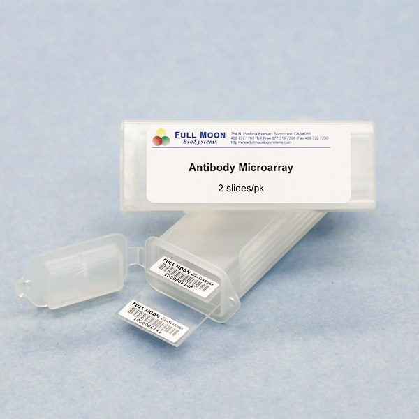
| Product Size: | 2 array slides per package for analyzing two samples (untreated vs. treated) |
| Featured Antibodies: | 1318 site-specific and phospho-specific antibodies; 2 replicates per antibody |
| Reactivity: | Human: 99% | Mouse: 92% | Rat: 66% |
| Suitable Sample Type: | Cell lysate | Tissue lysate |
| Detection Method: | Fluorescence | Compatible Scanners |
| Internal Controls: | beta-actin | GAPDH | Negative controls |
| Slide Dimensions: | 76 x 25 x 1 mm |
| Storage Condition: | 4°C for 6 months |
Product Details
14-3-3 theta/tau (Ser232), 14-3-3 zeta (Ser58), 14-3-3 zeta/delta (Ab-232), 14-3-3 zeta/delta (Thr232), 4E-BP1 (Ser65), 4E-BP1 (Thr36), 4E-BP1 (Thr45), 4E-BP1 (Thr70), Abl1 (Tyr204), Abl1 (Tyr412), ACC1 (Ser79), ACC1 (Ser80), ACK1 (Tyr284), AFX (Ser197), AKT (Ser473), AKT (Thr308), AKT (Tyr326), AKT1 (Ser124), AKT1 (Ser246), AKT1 (Thr450), AKT1 (Thr72), AKT1 (Tyr474), AKT2 (Ser474), ALK (Tyr1507), ALK (Tyr1604), AMPK1 (Thr174), Amyloid beta A4 (Thr743/668), Androgen Receptor (Ser213), Androgen Receptor (Ser650), A-RAF (Tyr301/302), Arrestin-1 (Ab-412), Arrestin-1 (Ser412), ASK1 (Ser83), ASK1 (Ser966), ATF-1 (Ser63), ATF2 (Ab-112/94), ATF2 (Ser112/94), ATF2 (Ser62/44), ATF2 (Thr69/51), ATF2 (Thr71/53), ATF2 (Thr73/55), ATF4 (Ser245), ATP1A1/Na+K+ ATPase1 (Ser23), ATPase (Ser16), ATP-Citrate Lyase (Ser454), ATRIP (Ser68/72), AurB (Thr232), AurB (Tyr12), Aurora Kinase (Thr288), AXL (Tyr691), and more.
Anerillas C, Herman A, Early SRC activation skews cell fate from apoptosis to senescence, Sci. Adv. 8 (14):eabm0756. DOI: 10.1126/sciadv.abm0756
Bernier M, Paul RK, Negative Regulation of STAT3-mediated Cellular Respiration by SirT1, J. Biol Chem, 2011, 286(22):19270-9
Chesnokova LS, Mosher B, Distinct early role of PTEN regulation during HCMV infection of monocytes. PNAS 2024 Mar 19;121(12):e2312290121. doi: 10.1073/pnas.2312290121
Chesnokova LS, Yurochko AD, Using a Phosphoproteomic Screen to Profile Early Changes During HCMV Infection of Human Monocytes. Methods Mol Biol. 2021;2244:233-246. doi: 10.1007/978-1-0716-1111-1_12. PMID: 33555590Dong S, Jia C, The REG gamma Proteasome Regulates Hepatic Lipid Metabolism through Inhibition of Autophagy, Cell Metabolism. 2013, 18(3), 380-391
Chicon-Bosch M, Sanchez-Serra S, Multi-omics profiling reveals key factors involved in Ewing sarcoma metastasis, Mol Oncol. 2025 Jan 5. doi: 10.1002/1878-0261.13788
Earwaker P, Anderson C, RAPTOR up-regulation contributes to resistance of renal cancer cells to PI3K-mTOR inhibition, PLoS ONE, 2018, 13(2): e0191890
Eke I, Hehlgans S, Comprehensive analysis of signal transduction in three-dimensional ECM-based tumor cell cultures, J Biol Methods 2015; 2(4): e31
Falcomata C, Bartehl S, Selective multi-kinase inhibition sensitizes mesenchymal pancreatic cancer to immune checkpoint blockade by remodeling the tumor microenvironment, Nat Cancer. 2022 Jan 31. doi: 10.1038/s43018-021-00326-1
Fuchs, C, Medici G, CDKL5 deficiency predisposes neurons to cell death through the deregulation of SMAD3 signaling, Brain Pathol. 2019 Feb 22. doi: 10.1111/bpa.12716
Gryshkova V, Cotter M, Phosphoprotein expression profiles in rat kidney injury: Source for potential mechanistic biomarkers, J Cell Mol Med, 2019 Jan 12. doi: 10.1111/jcmm.14103
Gu Y, Liu Y, Tumor-educated B cells selectively promote breast cancer lymph node metastasis by HSPA4-targeting IgG, Nat Med, 2019 Jan 14. doi: 10.1038/s41591-018-0309-y
Han Z, Yan Z, Targeting ABCD1-ACOX1-MET/IGF1R axis suppresses multiple myeloma. Leukemia. 2025. doi.org/10.1038/s41375-025-02522-9
He D, Wu H, Gut stem cell aging is driven by mTORC1 via a p38 MAPK-p53 pathway, Nat Commun. 2020 Jan 2; 11(1):37. doi: 10.1038/s41467-019-13911-x
Hillard T, Miklossy G, 15∝-methoxypuupehenol induces antitumor effects in vitro and in vivo against human glioblastoma and breast cancer models, Mol Cancer Ther, 2017 Jan 9, doi: 10.1158/1535-7163
Howell MC Jr, Green R, EGFR TKI resistance in lung cancer cells using RNA sequencing and analytical bioinformatics tools, J Biomol Struct Dyn. 2022 Dec 16;1-20. doi: 10.1080/07391102.2022.2153269
Hu C, Li L, Complement Inhibitor CRIg/FH Ameliorates Renal Ischemia Reperfusion Injury via Activation of PI3K/AKT Signaling, J Immunol, 2018 Nov 14, doi: 10.4049/jimmunol.1800987
Hwang HJ, Kang D, FLRT2 prevents endothelial cell senescence and vascular aging by regulating the ITGB4/mTORC2/p53 signaling pathway, JCI Insight. 2024 Apr 8;9(7):e172678. doi: 10.1172/jci.insight.172678
Jiang HL, Sun HF, SSBP1 Suppresses TGF-β-Driven Epithelial-to-Mesenchymal Transition and Metastasis in Triple-Negative Breast Cancer by Regulating Mitochondrial Retrograde Signaling, Cancer Res 2016 76(4):952-64
Jiang R, Smailovic U, Increased CSF-decorin predicts brain pathological changes driven by Alzheimer’s Aβ amyloidosis, Acta Neuropathol Commun. 2022 Jul 4;10(1):96. doi: 10.1186/s40478-022-01398-5
Koppenhagen P, Dickreuter E, Head and neck cancer cell radiosensitization upon dual targeting of c-Abl and beta1-integrin. Radiother Oncol. 2017 Jul;53:184-190
Lee PC, Fang YF, Targeting PKCδ as a Therapeutic Strategy against Heterogeneous Mechanisms of EGFR Inhibitor Resistance in EGFR-Mutant Lung Cancer, Cancer Cell, 2018 Dec 10;34(6):954-969.e4
Li L, Zhao D, REGγ deficiency promotes premature aging via the casein kinase 1 pathway, PNAS 2013, 110(27): 11005-11010
Liao W, Hu R,Oleic acid regulates CD4+ T cells differentiation by targeting ODC1-mediated STAT5A phosphorylation in Vogt-Koyanagi-Harada disease, Phytomed. 2025 March 18, https://doi.org/10.1016/j.phymed.2025.156660.
Lim HS, Sohn E, Ethanol Extract of Elaeagnus glabra f. oxyphylla Branches Alleviates the Inflammatory Response Through Suppression of Cyclin D3/Cyclin-Dependent Kinase 11p58 Coupled to Lipopolysaccharide-Activated BV-2 Microglia, Natural Product Comm. January 2022.Phospho Explorer Array doi:10.1177/1934578X221075079
Liu Y, Murazzi I, Sarcoma cells secrete hypoxia-modified collagen VI to weaken the lung endothelial barrier and promote metastasis, Cancer Res 2024. doi.org/10.1158/0008-5472.CAN-23-0910
Mitrovský O, Myslivcová D, Inhibition of casein kinase 2 induces cell death in tyrosine kinase inhibitor resistant chronic myelogenous leukemia cells, Plos One, May 4, 2023, https://doi.org/10.1371/journal.pone.0284876
Mulder SE, Dasgupta A, JNK signaling contributes to skeletal muscle wasting and protein turnover in pancreatic cancer cachexia, Cancer Letters. 2020 Jul 28. https://doi.org/10.1016/j.canlet.2020.07.025
Peaslee, C, Esteva‐Font, C, Doxycycline Significantly Enhances Induction of iPSCs to Endoderm by Enhancing survival via AKT Phosphorylation. Hepatology, 2021. Accepted Author Manuscript. https://doi.org/10.1002/hep.31898
Pellegrinelli V, Peirce VJ, Adipocyte-secreted BMP8b mediates adrenergic-induced remodeling of the neuro-vascular network in adipose tissue, Nat Commun, 2018 26;9(1):4974
Peran I, Dakshanamurthy S, Cadherin 11 Promotes Immunosuppression and Extracellular Matrix Deposition to Support Growth of Pancreatic Tumors and Resistance to Gemcitabine in Mice, Gastroenterology. 2021 Mar;160(4):1359-1372.e13
Pulito C, Sacconi A, Antibody Array as a Tool for Screening of Natural Agents in Cancer Chemoprevention, Methods Mol Biol, 2016 1379:189-99
Rebollo-Hermanz, Zhang Q, Cocoa Shell Aqueous Phenolic Extract Preserves Mitochondrial Function and Insulin Sensitivity by Attenuating Inflammation between Macrophages and Adipocytes In Vitro, Mol Nutr Food Res. 2019 May;63(10):e1801413. doi: 10.1002/mnfr.201801413
Sankar A, Cho HM, Antibody-drug conjugate αEGFR-E-P125A reduces triple-negative breast cancer vasculogenic mimicry, motility, and metastasis through inhibition of EGFR, integrin, and FAK/STAT3 signaling, Cancer Res Commun. 2024 Feb 5. doi: 10.1158/2767-9764.CRC-23-0278
Schatton, T, Itoh Y, Inhibition of melanoma cell-intrinsic Tim-3 stimulates MAPK-dependent tumorigenesis, Cancer Res. 2022 Aug 18;CAN-22-0970. doi: 10.1158/0008-5472.CAN-22-0970
Scott ML, Woodby BL, Human papillomavirus type 16 E5 inhibits interferon signaling and supports episomal viral maintenance, J Virol. 2019 Oct 30. doi: 10.1128/JVI.01582-19
Shi F, Guo H, The PI3K inhibitor GDC-0941 enhances radiosensitization and reduces chemoresistance to temozolomide in GBM cell lines, Neuroscience 2017, 346:298-308
Suh JS, Lee HJ, Control of cancer stem cell like population by intracellular target identification followed by the treatment with peptide-siRNA complex, Biochem & Biophys Res Commu, 2017 May 26, https://doi.org/10.1016/j.bbrc.2017.05.148
Tang N, Zhang J, PP2Acα inhibits PFKFB2-induced glycolysis to promote termination of liver regeneration, Biochem Biophys Res Commun. 2020 March 16. https://doi.org/10.1016/j.bbrc.2020.03.002
Vehlow A, Kapproth E, Interaction of Discoidin Domain Receptor 1 with a 14-3-3-Beclin-1-Akt1 Complex Modulates Glioblastoma Therapy Sensitivity, Cell Rep 2019; 26(13):3672-3683.e7
Venkatesh H, Tam Lydia, Targeting neuronal activity-regulated neuroligin-3 dependency in high-grade glioma, Nature, 2017 549:533–537
Xie X, Lv H, HBeAg mediates inflammatory functions of macrophages by TLR2 contributing to hepatic fibrosis, BMC Med 2021; 19:247. https://doi.org/10.1186/s12916-021-02085-3
Xu J, Zhou L, The REGγ-proteasome forms a regulatory circuit with IκBε and NFκB in experimental colitis, Nat Commun, 2016 Feb 7:10761
Vargas B, Boslett J, Mechanism by Which PF-3758309, a Pan Isoform Inhibitor of p21-Activated Kinases, Blocks Reactivation of HIV-1 Latency. Biomolecules. 2023; 13(1):100. https://doi.org/10.3390/biom13010100
Yin GN, Kim DK, Latrophilin-2 is a novel receptor of LRG1 that rescues vascular and neurological abnormalities and restores diabetic erectile function, Exp Mol Med. 2022 May 13. doi: 10.1038/s12276-022-00773-5
You KS, Yi, YW, Dual Inhibition of AKT and MEK Pathways Potentiates the Anti-Cancer Effect of Gefitinib in Triple-Negative Breast Cancer Cells. Cancers 2021. 13(6): 1205
Yoon N, Lee H, A Metabolomics Investigation of the Metabolic Changes of Raji B Lymphoma Cells Undergoing Apoptosis Induced by Zinc Ions, Metabolites 2021. 11(10): 689
Services
If you don’t have access to a microarray, send the finished arrays to our lab for scanning. Raw scan images are delivered in tiff format.
Cost: Free
Array Image Quantification and Analysis Service includes data extraction, data organization and analysis of the array images obtained through our array scanning service.
Cost: $350 per slide
Complete Antibody Array Assay Service allows investigators to send research samples to our laboratory for analysis. There is no need to purchase the arrays and reagents and running the assays yourself. Simply select the array of your choice, and then send off the samples to our lab. This convenient hands-off approach offers quick turnaround and reliable results, saving you valuable time and resources. All assays will be performed by our highly trained scientists at our headquarter in Sunnyvale, California. Results are delivered by email in 1-3 weeks.
Cost: $2,310 per sample


