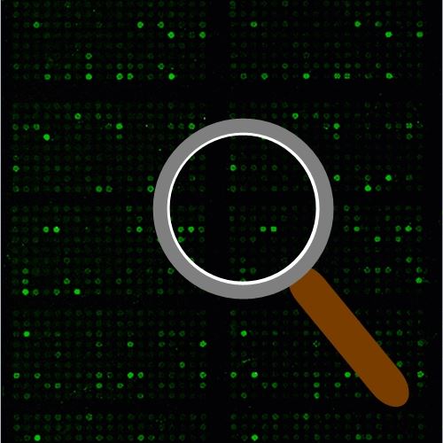
NFkB Phospho Antibody Array
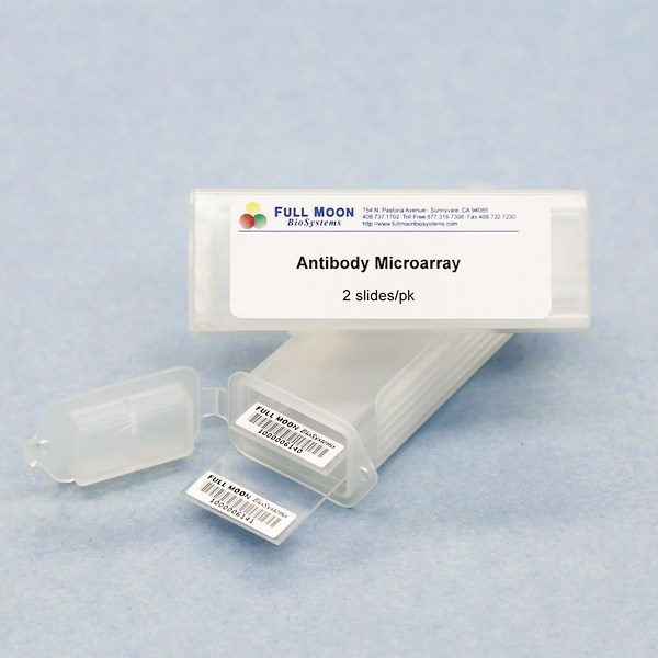
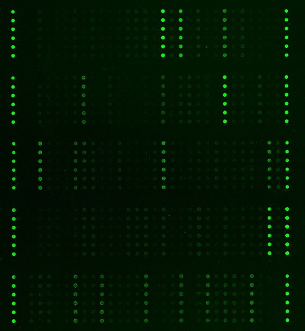
NFkB Phospho Antibody Array is a high-throughput ELISA based antibody array for qualitative protein phosphorylation profiling. It is suitable for comparing normal samples to treated or diseased samples, and identifying candidate biomarkers. This array features site-specific and phospho-specific antibodies for studying tyrosine phosphorylation and serine/threonine phosphorylation at specific sites.
Key Features
- Site-specific phosphorylation profiling and screening with 215 antibodies
- Antibodies covalently immobilized on 3D polymer coated glass slide
- Fluorescent detection
Specifications
| Product Size: | 2 array slides per package for two samples (untreated vs. treated) |
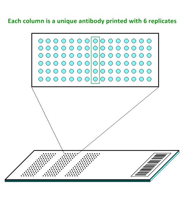
| Featured Antibodies: | 215 site-specific and phospho-specific antibodies; 6 replicates per antibody |
| Reactivity: | Human: 100% | Mouse: 94% | Rat: 55% |
| Suitable Sample Type: | Cell lysate | Tissue lysate |
| Detection Method: | Fluorescence | Compatible Scanners |
| Internal Controls: | Positive controls: beta-actin | GAPDH | Negative controls |
| Slide dimensions: | 76 x 25 x 1 mm |
| Storage Condition: | 4°C for 6 monthss |
Product Details
AKT (Ser473), AKT (Thr308), AKT (Tyr326), AKT1 (Ser124), AKT1 (Ser246), AKT1 (Thr450), AKT1 (Thr72), AKT1 (Tyr474), AKT1S1 (Thr246), AKT2 (Ser474), BLNK (Tyr84), BLNK (Tyr96), BTK (Tyr222), BTK (Tyr550), CK2-b (Ser209), COT (Thr290), Elk-1 (Ser383), Elk-1 (Ser389), Elk-1 (Thr417), GSK3a/b (Tyr216/279), GSK3b (Ser9), HDAC1 (Ser421), HDAC10, HDAC2 (Ser394), HDAC3 (Ser424), HDAC4 (Ser632), HDAC5 (Ser259), HDAC5 (Ser498), HDAC6 (Ser22), HDAC7, HDAC8 (Ser39), HDAC9, Histone H3.1 (Ser10), IkBaSer32/36), IkB-a (Tyr305), IkB-a (Tyr42), IkB-b (Ser23), IkB-b (Thr19), IkB-e (Ser22), IKKa (Thr23), IKKa/b (Ser180/181), IKKb (Tyr188), IKKb (Tyr199), IKKg (Ser31), IKKg (Ser85), JNK1/2/3 (Thr183/Tyr185), LCK (Ser59), LCK (Tyr192), LCK (Tyr393), LCK (Tyr504), MSK1 (Phospho-Ser376), MSK1 (Ser212), MSK1 (Ser360), MSK1 (Thr581), NFkB-p100 (Ser872), NFkB-p100/p52 (Ser865), NFkB-p100/p52 (Ser869), NFkB-p105 (Ser927), NFkB-p105/p50 (Ser337), NFkB-p105/p50 (Ser893), NFkB-p105/p50 (Ser907), NFkB-p105/p50 (Ser932), NFkB-p65 (Ser276), NFkB-p65 (Ser311), NFkB-p65 (Ser468), NFkB-p65 (Ser529), NFkB-p65 (Ser536), NFkB-p65 (Thr254), NFkB-p65 (Thr435), P38 MAPK (Thr180), P38 MAPK (Tyr182), P38 MAPK (Tyr322), PI3K-p85-a (Tyr607), PI3K-p85-a/g (Tyr467/Tyr199), PKA CAT (Thr197), PKC-a/b II (Thr638), PKC-b (Ser661), PKC theta (Ser676), PKC theta (Thr538), PKC zeta (Thr410), PKC zeta (Thr560), PKR (Thr446), PKR (Thr451), PLCG1 (Tyr1253), PLCG1 (Tyr771), PLCG1 (Tyr783), PLCG2 (Tyr1217), PLCG2 (Tyr753), Ras-GRF1 (Ser916), Rel (Ser503), RelB (Ser552), SAPK/JNK (Thr183), SAPK/JNK (Tyr185), SUMO-1, SUMO-2/3, SYK (Tyr323), SYK (Tyr348), SYK (Tyr525), TAK1 (Thr184), TGFBR1, TGFBR2, TNFR1, TNFR2, TRADD, Ubiquitin, ZAP-70 (Tyr292), ZAP-70 (Tyr315), Zap-70 (Tyr319), Zap-70 (Tyr493)
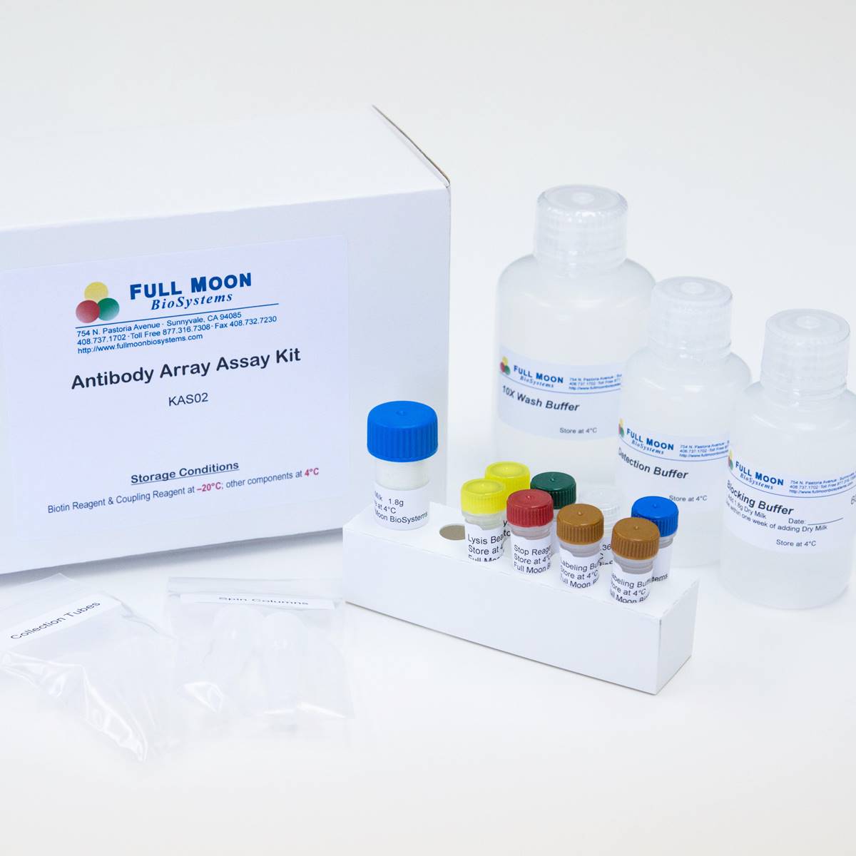
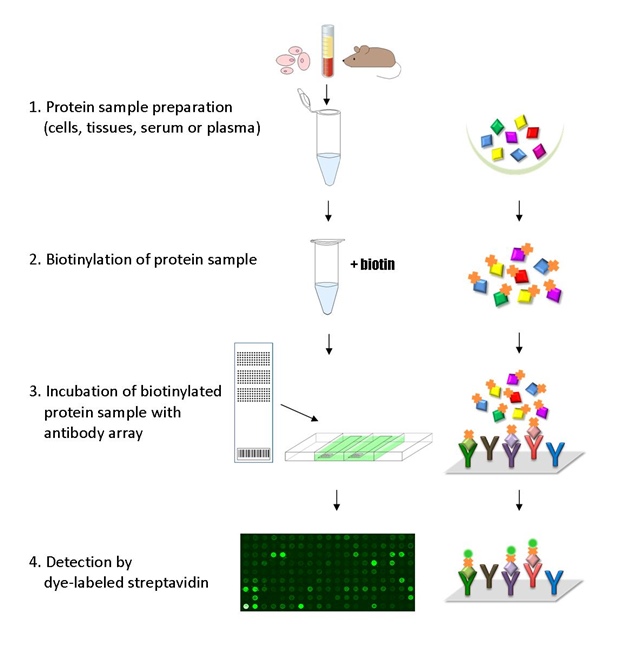
The ELISA based NF-KappaB Phospho Antibody Array platform involves four major steps:
- Protein extraction with non-denaturing lysis buffer
- Biotinylation of protein samples
- Incubation of labeled samples with antibody array
- Detection by dye conjugated streptavidin
![]() GAL File (To download, right click on the file name, then choose “Save target as”)
GAL File (To download, right click on the file name, then choose “Save target as”)
Bera A, Russ E, Proteomic Analysis of Inflammatory Biomarkers Associated With Breast Cancer Recurrence, Mil Med. 2020 Jan 7;185(Supplement_1):669-675. doi: 10.1093/milmed/usz254
Liu W, Li C, Cerebrospinal fluid of chronic osteoarthritic patients induced interleukin-6 release in human glial cell-line T98G, Anethesiol & Pain Med, 2019 Oct 8. doi: 10.21203/rs.2.15747/v1
Odeh A, Eddini H, Senescent Secretome of Blind Mole Rat Spalax Inhibits Malignant Behavior of Human Breast Cancer Cells Triggering Bystander Senescence and Targeting Inflammatory Response, Int. J. Mol. Sci. 2023. 24(6): 5132; https://doi.org/10.3390/ijms24065132
Shi Y, Yu J, RhTyrRS (Y341A), a novel human tyrosyl-tRNA synthetase mutant, stimulates thrombopoiesis through activation of the VEGF-R II/NF-κB pathway, Biochem Pharmacol. 2019 Sep 9:113634. doi: 10.1016/j.bcp.2019.113634
Singh MV, Wong T, BLNK/BTK: Novel Components in the NF-kappa B Pathways of Endothelial Cells in Diabetic Condition. J Endocr Soc. 2021;5(Suppl 1):A318-A319. Published 2021 May 3. doi:10.1210/jendso/bvab048.649
Srivastava M, Eidelman O, Serum Biomarkers for Racial Disparities in Breast Cancer Progression, Mil Med. 2019 Mar 1;184(Suppl 1):652-657. doi: 10.1093/milmed/usy417
Pan X, Jiang B, STC1 promotes cell apoptosis via NF-κB phospho-P65 Ser536 in cervical cancer cells, Oncotarget 2017; 8(28):46249-46261
Services
If you don’t have access to a microarray, send the finished arrays to our lab for scanning. Raw scan images are delivered in tiff format.
Cost: Free
Array Image Quantification and Analysis Service includes data extraction, data organization and analysis of the array images obtained through our array scanning service.
Cost: $250 per slide
Complete Antibody Array Assay Service allows investigators to send research samples to our laboratory for analysis. There is no need to purchase the arrays and reagents and running the assays yourself. Simply select the array of your choice, and then send off the samples to our lab. This convenient hands-off approach offers quick turnaround and reliable results, saving you valuable time and resources. All assays will be performed by our highly trained scientists at our headquarter in Sunnyvale, California. Results are delivered by email in 1-3 weeks.
Cost: $1,520 per sample

