Explorer Antibody Micorarray
Explorer Antibody Array is a high-throughput ELISA based antibody microarray for qualitative/semi-quantitative protein expression profiling and screening. The array is designed for comparing normal samples to treated or diseased samples, and identifying candidate biomarkers.
Key Features
- Qualitative/semi-quantitative protein expression profiling with 656 antibodies
- Antibodies covalently immobilized on 3D polymer coated glass slide
- Fluorescent detection
Specifications
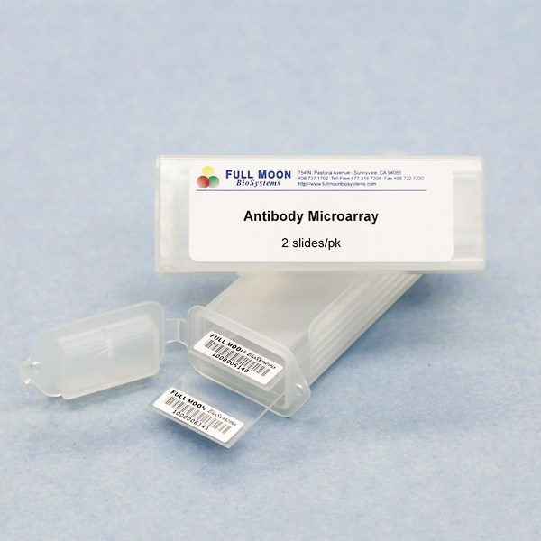
| Product Size: | 2 array slides per package for two samples (untreated vs. treated) |
| Featured Antibodies: | 656 antibodies; 2 replicates per antibody |
| Reactivity: | Human |
| Suitable Sample Type: | Cell lysate | Tissue lysate | Serum | Culture Media |
| Detection Method: | Fluorescence | Compatible Scanners |
| Internal Controls: | Positive controls: beta-actin | GAPDH | Negative controls |
| Slide dimensions: | 76 x 25 x 1 mm |
| Storage Condition: | 4°C for 6 months |
Product Details
Acinus, Actin Muscle Specific, Actin Pan, Actin skeletal muscle, Activin Receptor Type II, Adenovirus, Adenovirus Type 2 E1A, Adenovirus Type 5 E1A, ARF-6, AIF, Alkaline Phosphatase, Alpha Fetoprotein, Alpha Lactalbumin, alpha-1-antichymotrypsin, alpha-1-antitrypsin, Amphiregulin, Amylin Peptide, Amyloid A, Amyloid A4 Protein Precursor, Amyloid Beta, Androgen Receptor, Ang-1, Ang-2, APC, APC11, APC2, Apolipoprotein D, A-Raf, ARC, Ask1, ATM, Axonal Growth Cones, b Galactosidase, b-2-Microglobulin, B7-H2, BAG-1, Bak, Bax, B-Cell, B-cell Linker Protein, bcl-2a, bcl-6, bcl-10, bcl-X, bcl-XL, Bim, Biotin, Bonzo, Bovine Serum Albumin, BRCA2, BrdU, Bromodeoxyuridine, CA125, CA19-9, c-Abl, Cadherin Pan, Cadherin-E, Cadherin-P, Calcitonin, Calcium Pump ATPase, Caldesmon, Calmodulin, Calponin, Calretinin, Casein, Caspase 1, Caspase 2, Caspase 3, Caspase 5, Caspase 6, Caspase 7, Caspase 8, Caspase 9, Catenin alpha, Catenin beta, Catenin gamma, Cathepsin D, CCK-8, CD1, CD10, CD100/Leukocyte Semaphorin, CD1a, CD1b, CD2, CD3zeta, CD4, CD5, CD6, CD8, CD9 and more
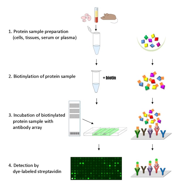
The ELISA based Explorer Antibody Array platform involves four major steps:
- Protein extraction with non-denaturing lysis buffer
- Biotinylation of protein samples
- Incubation of labeled samples with antibody array
- Detection by dye conjugated streptavidin

![]() GAL File (To download, right click on the file name, then choose “Save target as”)
GAL File (To download, right click on the file name, then choose “Save target as”)
Bagnis, A, Izzotti, A, Aqueous humor oxidative stress proteomic levels in primary open angle glaucoma, Experimental Eye Research, 2012, 103:55–62
Burcham PC, Raso A, Airborne Acrolein Induces Keratin-8 (Ser-73) Hyperphosphorylation and Intermediate Filament Ubiquitination in Bronchiolar Lung Cell Monolayers, Toxicology May 7 2014, 319:44-52
Guamanova NG, Vasilyev DK, Application of an antibody microarray for serum protein profiling of coronary artery stenosis, Biochem Biophys Res Commun, 2022, 631:55-63
, Visible red and infrared light alters gene expression in human marrow stromal fibroblast cells, Orthodontics & Craniofacial Research 2015; 18(Suppl.1): 50–61
Fan Q, Zhou J, Chip-based serum proteomics approach to reveal the potential protein markers in the sub-acute stroke patients receiving the treatment of Ginkgo Diterpene Lactone Meglumine Injection, J Ethnopharmacol. 2020 May 12;112964. doi: 10.1016/j.jep.2020.11296
Klimushina MV, Gumanova NG, Direct labeling of serum proteins by fluorescent dye for antibody microarray, Biochem Biophys Res Commun 2017; 486(3):824-826
Lee MS, Kim JH, Prognostic Significance of CREB-Binding Protein and CD81 Expression in Primary High Grade Non-Muscle Invasive Bladder Cancer: Identification of Novel Biomarkers for Bladder Cancer Using Antibody Microarray, PLOS One, 2015 10(4):e0125405
Moon, KM, Park, Y, The Effect of Secretory Factors of Adipose-Derived Stem Cells on Human Keratinocytes, Intl J of Mol Sci 2012, 13(1):1239-1257
Quistad SD, Stotland A, Evolution of TNF-induced Apoptosis Reveals 550 My of Functional Conservation, PNAS 2014 111(26) 9567-9572
Ravishankar P, Dail A, Tears as the Next Diagnostic Biofluid: A Comparative Study between Ocular Fluid and Blood, Appl. Sci. 2022. 12(6):2884; https://doi.org/10.3390/app12062884
Siddique A, Ebrahim H, (−)-Oleocanthal Combined with Lapatinib Treatment Synergized against HER-2 Positive Breast Cancer In Vitro and In Vivo, Nutrients 2019; 11(2): 412
Yang T, Yao H, Effects of Lovastatin on MDA-MB-231 Breast Cancer Cells: An Antibody Microarray Analysis, Journal of Cancer, 2016; 7(2): 192-199
Yin SJ, Park D, An RNA interference based study for the role of ALDH1 in keratinocytes: DNA microarray, antibody–chip array and bioinformatics approaches, Process Biochemistry, 2014 49(10): 1612–1621
Zupancic K, Blejec A, Identification of plasma biomarker candidates in glioblastoma using an antibody-array-based proteomic approach, Radiology and Oncology, 2014, 48(3), 257–266
Services
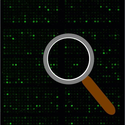
If you don’t have access to a microarray, send the finished arrays to our lab for scanning. Raw scan images are delivered in tiff format.
Cost: Free
Array Image Quantification and Analysis Service includes data extraction, data organization and analysis of the array images obtained through our array scanning service.
Cost: $250 per slide
Complete Antibody Array Assay Service allows investigators to send research samples to our laboratory for analysis. There is no need to purchase the arrays and reagents and running the assays yourself. Simply select the array of your choice, and then send off the samples to our lab. This convenient hands-off approach offers quick turnaround and reliable results, saving you valuable time and resources. All assays will be performed by our highly trained scientists at our headquarter in Sunnyvale, California. Results are delivered by email in 1-3 weeks.
Cost: $1,730 per sample


