Cell Cycle Control Phospho Antibody Array
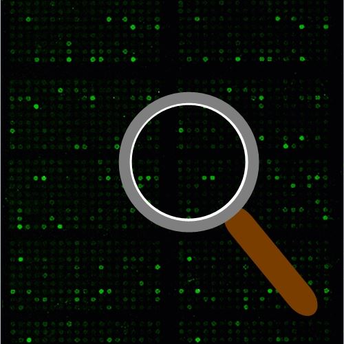
Cell Cycle Control Antibody Array is a high-throughput ELISA based antibody array for qualitative protein phosphorylation profiling. It’s designed for comparing normal samples to treated or diseased samples, and identifying candidate biomarkers. This array features site-specific and phospho-specific antibodies, allowing researchers to study tyrosine, serine, and threonine phosphorylation at specific sites.
Key Features
- Site-specific phosphorylation profiling and screening with 238 antibodies
- Antibodies covalently immobilized on 3D polymer coated glass slide
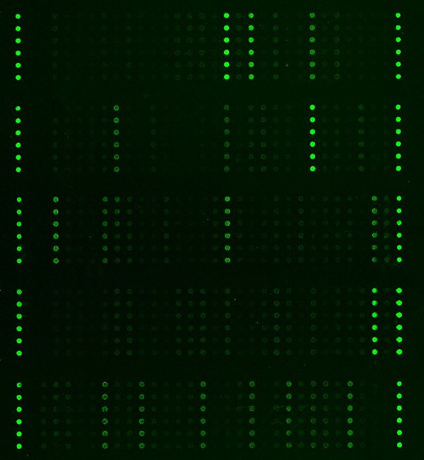
- Fluorescent detection
Specifications
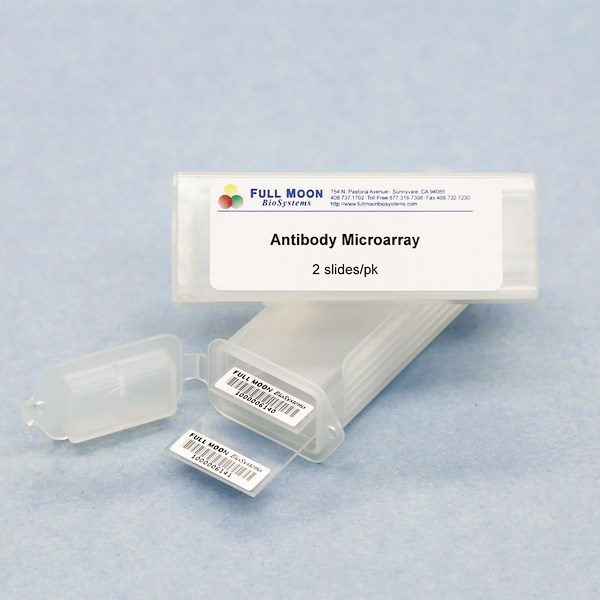
| Product Size: | 2 array slides per package for analyzing two samples (untreated vs. treated) |
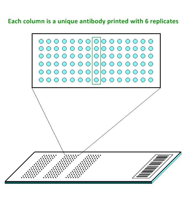
| Featured Antibodies: | 238 site-specific and phospho-specific antibodies; 6 replicates per antibody |
| Reactivity: | Human: 100% | Mouse: 85% | Rat: 57% |
| Suitable Sample Type: | Cell lysate | Tissue lysate |
| Detection Method: | Fluorescence | Compatible Scanners |
| Internal Controls: | beta-actin | GAPDH | Negative controls |
| Slide dimensions: | 76 x 25 x 1 mm |
| Storage Condition: | 4°C for 6 months |
Product Details
14-3-3 theta/tau (Ser232), 14-3-3 zeta (Ser58), 14-3-3 zeta/delta (Thr232), Abl1 (Thr754/735), Abl1 (Tyr204), Abl1 (Tyr412), AKT (Ser473), AKT (Thr308), AKT (Tyr326), AKT1 (Ser124), AKT1 (Ser246), AKT1 (Thr450), AKT1 (Thr72), AKT1 (Tyr474), AKT2 (Ser474), BRCA1 (Ser1423), BRCA1 (Ser1457), BRCA1 (Ser1524), c-Abl (Tyr245), c-Abl (Tyr412), CDC2 (Tyr15), CDC25A (Ser124), CDC25A (Ser75), CDC25B (Ser323), CDC25B (Ser353), CDC25C (Ser216), CDC25C (Thr48), CDK1/CDC2 (Thr14), CDK2 (Thr160), CDK7 (Thr170), Chk1 (Ser280), Chk1 (Ser286), Chk1 (Ser296), Chk1 (Ser301), Chk1 (Ser317), Chk1 (Ser345), Chk2 (Ser516), Chk2 (Thr383), Chk2 (Thr387), Chk2 (Thr68), Cyclin A (A1/A2), Cyclin A1, Cyclin B1 (Ser126), Cyclin B1 (Ser147), Cyclin D1 (Thr286), Cyclin D3 (Thr283), Cyclin E1 (Thr395), Cyclin E1 (Thr77), DNA-PK (Thr2638), DNA-PK (Thr2647), E2F1 (Thr433), E2F2, E2F4, E2F6, FKHR (Ser256), FKHR (Ser319), FKHRL1 (Ser253), FOXO1/3/4-PAN (Thr24/32), FOXO1A (Ser329), FOXO1A/3A (Ser322/325), GSK3a-b (Tyr216/279), GSK3beta (Ser9), HDAC1 (Ser421), HDAC2 (Ser394), HDAC3 (Ser424), HDAC4 (Ser632), HDAC5 (Ser259), HDAC5 (Ser498), HDAC6 (Ser22), HDAC7, HDAC8 (Ser39), HDAC9, HDAC10, Histone H2A.X (Ser139), MDM2 (Ser166), MDM4 (Ser367), Myc (Ser373), Myc (Ser62), Myc (Thr358), Myc (Thr58), p15INK, p18INK, p21Cip1 (Thr145), p27Kip1 (Ser10), p27Kip1 (Thr187), p300, p300/CBP, p53 (Ser6), p53 (Ser9), p53 (Ser20), p53 (Ser15), p53 (Ser33), p53 (Ser37), p53 (Ser46), p53 (Ser315), p53 (Ser366), p53 (Ser378), p53 (Ser392), p53 (Thr18), p53 (Thr81), p90RSK (Ab-359/363), p90RSK (Ser380), p90RSK (Thr359/Ser363), p90RSK (Thr573), p95/NBS1 (Ser343), PP2A-a (Tyr307), RAD51 (Tyr315), RAD52 (Tyr104), Rb (Ser608), Rb (Ser780), Rb (Ser795), Rb (Ser807), Rb (Ser811), Rb (Thr821), Smad2/3 (Thr8), Smad3 (Ser204), Smad3 (Ser208), Smad3 (Ser213), Smad3 (Ser425), Smad3 (Thr179), Smad4, TGFB1, TGFB2, TGFB3, TGFGBR2, TOP2A (Ser1106), WEE1 (Ser642)
Elias D, Vever H, Gene expression profiling identifies FYN as an important molecule in tamoxifen resistance and a predictor of early recurrence in patients treated with endocrine therapy, Oncogene 2014, 1-9
Gruosso T, Mieulet V, Chronic oxidative stress promotes H2AX protein degradation and enhances chemosensitivity in breast cancer patients, EMBO Mol Med 2016 Mar 22. pii: e201505891. doi: 10.15252/emmm.201505891
Lee S, Jang J, Latent Kaposi’s sarcoma-associated herpesvirus infection in bladder cancer cells promotes drug resistance by reducing reactive oxygen species, J Microbiol 2016 Nov, 4(11):782-788
Ma J, Sun F, Depletion of intermediate filament protein Nestin, a target of microRNA-940, suppresses tumorigenesis by inducing spontaneous DNA damage accumulation in human nasopharyngeal carcinoma, Cell Death and Disease 2014, 5:e1377
Services
If you don’t have access to a microarray, send the finished arrays to our lab for scanning. Raw scan images are delivered in tiff format.
Cost: Free
Array Image Quantification and Analysis Service includes data extraction, data organization and analysis of the array images obtained through our array scanning service.
Cost: $250 per slide
Complete Antibody Array Assay Service allows investigators to send research samples to our laboratory for analysis. There is no need to purchase the arrays and reagents and running the assays yourself. Simply select the array of your choice, and then send off the samples to our lab. This convenient hands-off approach offers quick turnaround and reliable results, saving you valuable time and resources. All assays will be performed by our highly trained scientists at our headquarter in Sunnyvale, California. Results are delivered by email in 1-3 weeks.
Cost: $1,520 per sample

