Cancer BioMarker Antibody Array
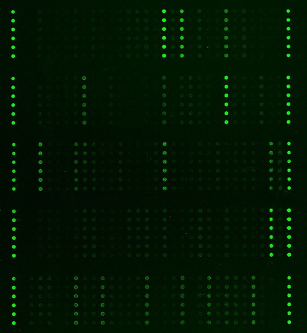
Cancer BioMarker Antibody Array is a high-throughput ELISA based antibody array designed for qualitative/semi-quantitative protein expression profiling, comparing normal samples to treated or diseased samples, and identifying candidate biomarkers. This array is specifically designed to detect changes in important markers associated with various cancer types.
Key Features
- Qualitative/semi-quantitative protein expression profiling with 247 antibodies
- Antibodies covalently immobilized on 3D polymer coated glass slide
- Fluorescent detection
Specifications
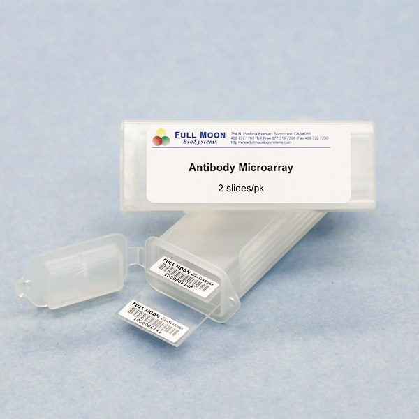
| Product Size: | 2 array slides per package for two samples (untreated vs. treated) |
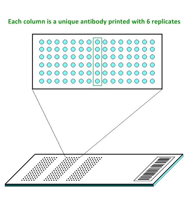
| Featured Antibodies: | 247 cancer biomarker related antibodies; 6 replicates per antibody |
| Reactivity: | Human |
| Suitable Sample Type: | Cell lysate | Tissue lysate | Serum | Culture Media |
| Detection Method: | Fluorescence | Compatible Scanners |
| Internal Controls: | Positive controls: beta-actin | GAPDH | Negative controls |
| Slide dimensions: | 76 x 25 x 1 mm |
| Storage Condition: | 4°C for 6 months |
PRODUCT DETAILS
ACT, ACTH, Adipolean Variant, Adiponectin, AFP, Androgen Receptor, Ang-1, Ang-2, APLP2, ApoE3, ApoF, ApoL1, ApoL2, Beta-2-Microglobulin, beta-NGF, BDNF, CA15-3, CA19-9, CA125, Cadherin-pan, CEA, Collagen I, Collagen II, Collagen III, Collagen IV, CRP, Death Receptor 5, Desmin, DNA mismatch repair protein Mlh1, DNA mismatch repair protein Mlh3, DNA mismatch repair protein Msh2, DNA mismatch repair protein Msh3, DNA mismatch repair protein MSH6, EGF, EGF Receptor, Endoglin(CD105), Eotaxin, Eotaxin-2, Eotaxin-3, Epithelial Cell Adhesion Molecule, E-Selectin, Estrogen Receptor, FAS, Ferritin, Fibroblast Growth Factor acidic (FGF-acidic), Fibroblast Growth Factor basic (FGF-basic), Fibronectin, Flt-1 (VEGFR1), Follicle Stimulating Hormone (FSH), Follistatin, Fractalkine, Free PSA, Granulocyte Colony-Stimulating Factor (G-CSF), Granulocyte/Macrophage Colony-Stimulating Factor (GM-CSF), Granzyme B, and more
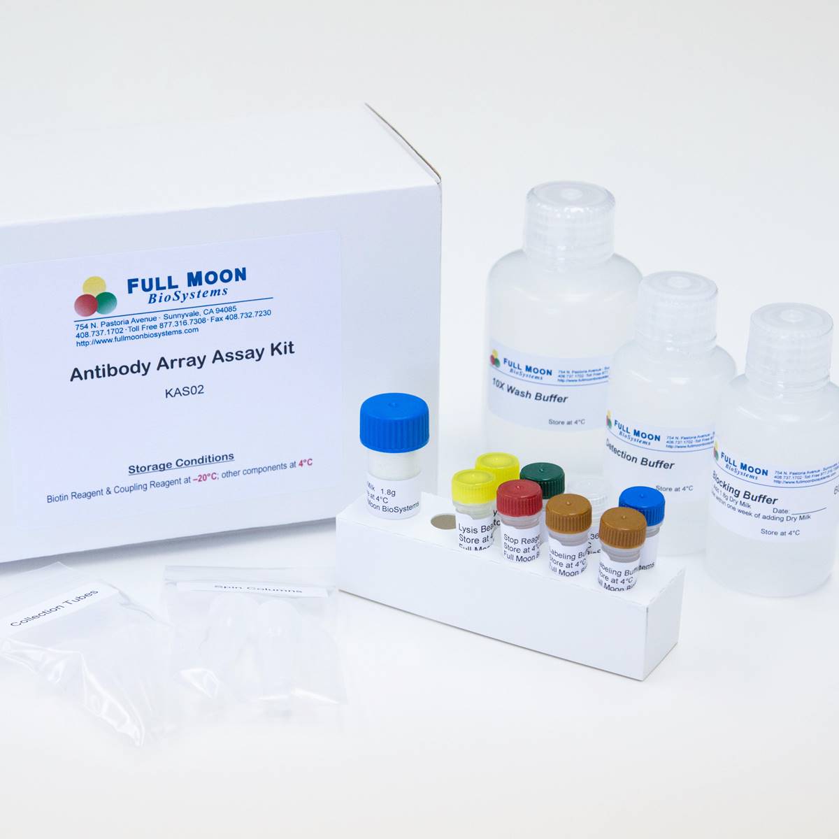
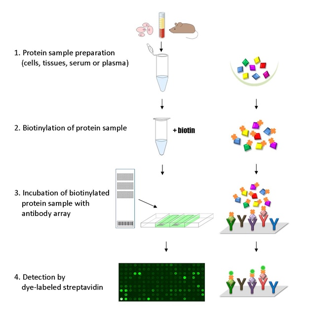
The ELISA based Cancer BioMarker Antibody Array platform involves four major steps:
- Protein extraction with non-denaturing lysis buffer
- Biotinylation of protein samples
- Incubation of labeled samples with antibody array
- Detection by dye conjugated streptavidin
![]() GAL File (To download, right click on the file name, then choose “Save target as”)
GAL File (To download, right click on the file name, then choose “Save target as”)
Hannafon B, Gin A, Metastasis-associated protein 1 (MTA1) is transferred by exosomes and contributes to the regulation of hypoxia and estrogen signaling in breast cancer cells, MoCell Communication and Signaling 2019 17:13. https://doi.org/10.1186/s12964-019-0325-7
Ravishankar P, Dail A, Tears as the Next Diagnostic Biofluid: A Comparative Study between Ocular Fluid and Blood, Appl. Sci. 2022. 12(6):2884; https://doi.org/10.3390/app12062884
ADDITIONAL SERVICES
If you don’t have access to a microarray, send the finished arrays to our lab for scanning. Raw scan images are delivered in tiff format.
Cost: Free
Array Image Quantification and Analysis Service includes data extraction, data organization and analysis of the array images obtained through our array scanning service.
Cost: $250 per slide
Complete Antibody Array Assay Service allows investigators to send research samples to our laboratory for analysis. There is no need to purchase the arrays and reagents and running the assays yourself. Simply select the array of your choice, and then send off the samples to our lab. This convenient hands-off approach offers quick turnaround and reliable results, saving you valuable time and resources. All assays will be performed by our highly trained scientists at our headquarter in Sunnyvale, California. Results are delivered by email in 1-3 weeks.
Cost: $1,520 per sample

