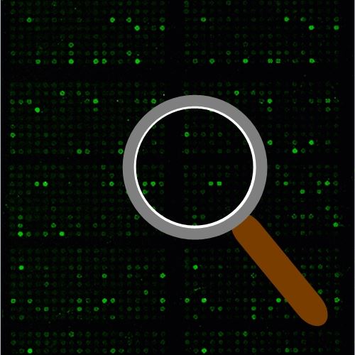
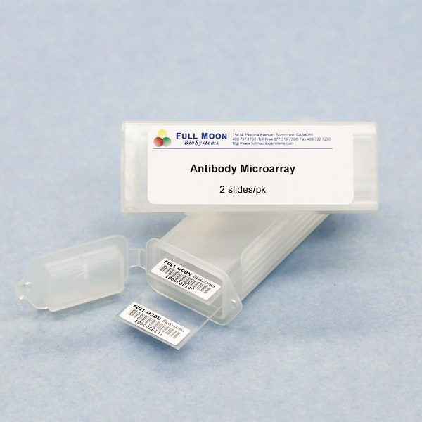
The Array Image Quantification and Analysis Service includes data extraction from raw images, data organization and analysis:
- Signal intensity for each spot
- Average signal intensity for replicate spots
- Coefficient of variation for replicate spots
- Ratios of phospho-specific antibody’s signal to site-specific antibody’s signal
- Fold change between control sample and treated sample
Cost and Deliverables
- Cost: $200/slide
- Deliverable: Array data in Excel format – Sample Analysis Results
- Turnaround Time: 2-4 days
- Delivery Method: Email
How to Use the Service
- Purchase any Full Moon BioSystems’ antibody arrays
- Complete assays using Cy3 or Cy5 labeled streptavidin (or equivalents)
- Send the arrays to Full Moon BioSystems for scanning
- Place an order for Image Analysis Service. Ordering options:
Preparing Slides For Scanning and Image Analysis
- Dry the slides according to the instructions in the Antibody Array User’s Guide.
- Place the slides in the original slide holder or a slide box.
- When using transparent holders, cover the slide holder with aluminum foil to protect the slides from light.
- Complete the Slide Submission Form and include it in the package.
- Send the package at room temperature within 1 week after completing the assay.
Slide Submission Form: PDF WORD
Shipping address
Attn: Array Scanning Service
Full Moon BioSystems, Inc.
754 North Pastoria Avenue
Sunnyvale, CA 94085
United States
Tel: 408.737.2875
Email:


